The body of the mushroom is called. What is the fruiting body of a mushroom? Types, development and structure of fruiting bodies
Body structure. The body of the mushroom is mycelium (mycelium)– an extensive network of thin threads – gif. The mycelium develops on the surface or inside the substrate and has greater contact with it, which ensures osmotic absorption of nutrients.
If the hyphae are divided by partitions (septa) into individual cells, then they form a cellular (septate) mycelium, if they represent one branched cell - non-cellular mycelium. The septum develops from the wall of the hypha to its center, where a pore remains through which the cytoplasm (as well as individual organelles) can move from one cell to another. Mycelium hyphae can be tightly intertwined (formation of sclerotia), forming false tissue - plectenchyma, from which the so-called fruiting body is formed (it differs from the real tissue of higher plants, formed by cell division in all directions). Sclerotia cells are rich in nutrients and help the fungus tolerate unfavorable conditions. From the sclerotia, mycelium or reproductive organs again develop. When connecting, the hyphae form cord-like strands - rhizomorphs. They partly perform a conducting function. The hyphae of their outer layers have thickened, often dark-colored walls and perform protective functions.
The hyphal wall contains up to 80-90% polysaccharides associated with proteins and lipids. The skeletal components of the wall (microfibrils) consist of chitin or cellulose (rarely). Beneath the cell wall is the protoplast. In the cytoplasm of fungal cells, plasmalemma and tonoplast, ribosomes, mitochondria, Golgi apparatus, endoplasmic reticulum and nucleus (nuclei) with a double membrane are clearly distinguishable. Between the cell wall and the plasma membrane are lomosomes, membrane structures that look like vesicles. The Golgi apparatus is not an essential structure of the cytoplasm. Consists of single cisternae and Golgi vesicles. Mitochondria with lamellar cristae, like in animals. An important feature of the structure of fungi is the absence of plastids. The kernels are very small. Spare products are stored in the form of glycogen; starch is never formed. Hyphae grow at their tips (apical growth)
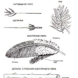
Structure of the fruiting body. The fruiting body of cap mushrooms usually consists of a cap and a stalk. At a young age, the cap is usually round, convex, and ovoid. With this shape, the fruiting body breaks through more easily to the surface of the soil or other substrate. hat different types mushrooms have sizes. The shape of the cap is also varied and is one of the important features in the taxonomy of mushrooms (conical, bell-shaped, convex).
Hymenophore. On the lower surface of the cap, tubes or plates are densely located; some species have spines or needles, similar to the spines of a hedgehog. These formations are called hymenophore. The inner surface of the tubes and the outer surface of the plates and spines are covered with a spore-bearing layer (hymenia). The tubes are arranged vertically, with their upper ends attached to the lower surface of the cap pulp, and the lower end of each tube has a hole (pore), through which the spores spill out and are dispersed by air currents. Spores are carried over long distances. Tubule pores in different species
mushrooms have characteristics in shape and size, and their edges have characteristics in color. The length of the tubes is also different. In addition, the tubular layer in some species is easily separated from the flesh of the cap,
while in others it does not separate, and this is an important systematic feature.
Features of records. The length and relative position of the plates on the lower surface of the cap are important for identifying mushrooms. In some types of mushrooms, all the plates are the same length (main plates), in others, the main plates do not reach the edge of the cap, and in this case the edge of the cap is called sterile, because there are no spores on it. Some types of mushrooms have forked plates.
General and private covers. General blanket (Velum)- this is a filmy formation that completely covers the fruiting body at the beginning of its development. As the fruiting body grows, the general covering usually breaks at the apex. In some types of mushrooms, scraps of the common cover remain on the surface of the cap in the form of scales or warts. They remain on the cap or quickly disappear. The lower part of the common cover remains at the base of the leg: in some species-shaped cup or bag-shaped and is called Volvo, in others, the remainder of the common veil is located in the form of rings, scales, or some other shape, or disappears, leaving barely noticeable traces. Private bedspread may be: filmy or cobwebby, covering the lower surface of the cap at a young age. It plays a protective role for developing spores. With the growth of the fruiting body, when the spores ripen, the private cover is torn off from the edge of the cap and remains on the stem in the form of a ring.
Often mushrooms enter into cohabitation with algae and even higher plants. In the process of symbiosis, new formations arise, for example, lichens, mycorrhiza (ectomycorrhiza and endomycorrhiza).
Reproduction. Fungi are characterized by vegetative, asexual and sexual reproduction.
I) Vegetative propagation(fragmentation):
- oidia - are formed as a result of the disintegration of mycelial hyphae into individual short cells, which can give rise to new mycelium (mucoraceae).
- chlamydospores - hyphae are formed in some places, are distinguished by a denser shell, and are dark-colored. They have a supply of nutrients inside. In addition to reproduction, they also perform the function of tolerating extreme conditions (increasing and decreasing temperature, humidity, etc.);
- In most yeasts, reproduction occurs by budding.
II. Asexual reproduction. With the help of spores. Endogenous disputes – inside special cells - sporangiospores. They are formed on special hyphae - sporangiophores. Exogenous spores – are called conidia. They are formed on special hyphae - conidiophores. Can form in airborne environments. Capable of budding and as a result chains of conidia are formed.
III. Sexual reproduction.
1. Gametogamy(isogamy, heterogamy) - two gametes merge (one or both are mobile, identical or of different sizes). Gametogamy is divided into isogamy, in which both gametes, mobile and morphologically indistinguishable, merge (copulate), and heterogamy(anisogamy), in which motile gametes differing in size and often in the degree of mobility merge (copulate) (for example, in representatives of the Chytridiomycota divisions).
2. Oogamy- reproductive structures are formed – oogonium and antheridium. IN oogonia an immobile large egg is formed, and in antheridia Small, motile sperm are formed, which eventually penetrate the egg and fertilize it. As a result, zygote (oospore).
3. Gametangiogamy(zygogamy) - the contents of two specialized reproductive structures - gametangia, not differentiated into gametes, merge. Gametangia are usually multinucleated, and as a result of their fusion, along with the fusion of the cytoplasm, multiple nuclear fusions occur.
4. Somatogamy- a process in which reproductive structures are not formed, but ordinary somatic or vegetative cells of mycelial hyphae merge. This type of sexual process is characteristic of some representatives of chytridiomycotes and hyphochytridiomycotes with a unicellular thallus. In this case, two single-celled individuals merge (chologamia).
The life cycle alternates:
In lower fungi: 1) diploid (zygote) and 2) haploid (mycelium).
U higher mushrooms: 1) haploid (n); 2) dikaryon (n+n); 3) diploid (2n).
The main stages of the sexual process in fungi.
1. Plasmogamy- fusion of the cytoplasm of two sexually specialized cells, the transition of the nucleus and cytoplasm into the female reproductive structure or even into a somatic cell.
2. Karyogamy– nuclear fusion and, as a result, diploidization. In lower fungi (zygomycetes, endomycetes in marsupial fungi), nuclear fusion occurs immediately after plasmogamy. In higher fungi, this process is delayed and occurs in dikaryotic cells, often after the formation of the corresponding morphological structures - fruiting bodies.
3. Meiosis (reduction division) typically follows karyogamy. Then one or more mitotic divisions often occur. Ultimately, the number of spores is most often 4 (2).
Sexual relationship of mushrooms:
Heterothallism(dioecious) and homothallism(bisexual).
Alternative classification (Whittaker)
- Monera (prokaryotes)
- Protista (unicellular colonial eukaryotes)
- Multicellular eukaryotes – Plantae, Fungi, Animalia.
Classification.
- Empire Opisthocontae (postoflagellates)
- Superkingdom Eucariota (nuclear organisms)
- Kingdom of Fungi
- Subkingdom Mucobionta
Kira Stoletova
For a mushroom picker, you need to know the structure of mushrooms. This will help distinguish edible species from inedible. The fruiting body of the fungus is formed by intertwining mycelial hyphae - thin threads tightly adjacent to each other. In the specialized part, spores are formed as a result of the sexual process. Has different sizes and shapes.

General structure
The mushroom kingdom is huge. Its representatives often differ significantly from each other. Majority known species belong to the order Agariaceae. This includes honey mushrooms, chanterelles, saffron milk caps, champignons and others.
The body of the fungus consists of hyphae. Translated from Greek, this term means “web”, “fabric”. These threads are formed by cells with strong walls. Characterized by apical growth. Branched, may not have partitions and often consist of one large cell with nuclei (in lower ones).
Vegetative part
Irina Selyutina (Biologist):
Indeed, the hyphae from which the mycelium is formed have apical growth and branch abundantly. The closer they are located to the growing top, the younger their “branches” are. During the formation of sporulation organs, and often in vegetative organs, fungal threads are tightly intertwined and form plectenchyma.
The parallel connection of hyphae forms mycelial strands, which are very visible at the base of large fruiting bodies. Water and nutrients flow through them.
Often branched hyphae, meeting small roots of trees, entwine them like a cover. Sometimes they penetrate into their cells and form balls of hyphae there. Swellings and root branches appear. This symbiosis of the fungal body with the plant is called mycorrhiza or fungal root: thanks to this, the fungi receive glucose from the plants, and the plants do not need water and minerals.
Fruit part
Representatives of the mushroom kingdom reproduce in 2 ways:
- vegetatively - pieces of mycelium;
- with the help of spores - an asexual and hollow process.
The fruiting body of the mushroom is the reproductive part, scientifically called sporocarp or carpophorus.
It is formed by some specimens during sporulation. It consists of intertwined threads that form the pulp - the actual body mass. This is a false tissue called plectenchyma. The function of a separate part of it is the formation of spores. To examine them, the cap is placed on a sheet of paper. After some time, a gray coating will appear on it - this is spores. When placed in a favorable environment, they germinate. A mycelium forms, and eventually a mushroom.
Irina Selyutina (Biologist):
Plectenchyma, or false parenchyma, differs from real tissue in its origin. False tissue of fungi is formed by interlacing threads of mycelium, while in higher plants tissues are formed by cell division in all directions. Under a microscope, plectenchyma often resembles ordinary parenchyma (the main tissue of plants), and sometimes it may even contain differentiation of covering, conducting, etc.
The fruiting body of ascomycete mushrooms is called ascocarp or ascoma. In basidiomycetes there is a basidiocarp or basidiome.
Types of fruiting bodies
Representatives of the mushroom kingdom have different shapes.
In many sources there is a division into groups:
- hat-footed;
- sessile - in the form of growths, hooves, cantilever-shaped (they lack a leg or stump, hence the name);
- round, pear-shaped, etc.;
- prostrate, lobed, in the form of corals, ear, star, etc.
The shape of the mushroom's fruiting body may change during growth. There are species that look like a star and acquire a phallic or lobed resemblance.
Structure of the fruiting body
Mushrooms are divided into lower and higher according to structural features. The first group includes mold species. Their spores are always in the air and, under favorable conditions, begin to multiply. Many representatives are present in soil and manure. Hyphae appear on bread plant products. They entwine them like a web. Spores are produced in sporangia that form at the ends of hyphae.
In most cases, the fruiting body of agaric mushrooms consists of a cap and a stalk. They come in different colors and sizes. But in some specimens these organs are weakly expressed or absent altogether.
hat

Its components, like those of the vegetative bodies of fungi, are hyphae. The surface of the cap is formed by covering threads - this is the skin. Its function is to prevent the evaporation of moisture from internal tissues. It can be dry or slimy, depending on the weather. In many species it is easily separated from the pulp over the entire surface, in others it can only be removed from the edges. There are specimens with firmly attached skin.
The pulp of this part of the mushroom's body is sterile tissue. It can be loose or elastic. In many species it is brittle. It has a different smell: light, pleasant or sharp, burning. In most cases the color is light. May change with age. When cut, it remains the same or becomes darker. In many specimens, some of the hyphae have thickened walls. Inside they have juice, which is often called milky. In some representatives it is present in abundant quantities, in others - in small quantities. It can be colorless or colored.
The shape of the caps is different:
- cushion-shaped;
- spherical;
- funnel-shaped;
- bell-shaped, etc.
The edges can be turned up or down. Some specimens change the shape of the cap throughout life cycle.
Depending on the type of hymenophore located in this part of the fruiting body, the mushroom is called:
- Lamellar: Bottom part caps are represented by plates. These include, for example, moss mushroom, boletus mushroom, and oiler.
- Tubular: the hymenophore is represented by tubes. This group includes mushrooms such as saffron milk cap, champignons, and autumn honey fungus.
- Marsupial: has a cellular or sinuous-folded surface. The group includes truffle, morel, and common string.
Hymenophore
It is part of the structure of the fruiting body of the cap mushroom. This formation is a sign of high organization. Specimens with a hymenophore have a thin spore-bearing layer on their surface - the hymenium. It consists of microscopic cells - basidium. In the first case, it fuses with the leg, in the second, it does not reach it. Sometimes he runs down (goes down) along it. Can be easily separated from the cap or tightly attached.
Hymenophore species:
- smooth;
- folded;
- spiny;
- tubular;
- labyrinthine;
- lamellar.
It has great importance for the reproduction process. When fruiting cells are dispersed from the hymenium, a new vegetative body of the fungus is formed under appropriate environmental conditions.
Controversy
The spores are visible only with a microscope. Compound:
- cytoplasm;
- shell – smooth, spinous, bristly or warty;
- core;
- other organelles.
Dimensions are 10-25 microns. The shape is varied: round, oval, granular, star-shaped, spindle-shaped. In the external environment they look like powder, so in mycology there is a special term - spore powder. They can be transparent or colored. Mycologists place great importance on these characteristic features, because they help to accurately determine the species of a specimen.
Leg

The stem (stump), like the cap, is a part (organ) of the fruiting body of the mushroom. It functions as a support, so the hyphae consist of huge, durable cells. They are distinguished by a characteristic thickened shell. The hyphae are arranged from bottom to top, collected in bundles and placed in parallel. They serve to deliver water and nutrients from the vegetative body of the mushroom to the cap.
The following types of legs are found:
- hollow;
- solid;
- mixed - the outer part is dense, the inner part is spongy.
They also differ in shape and thickness. They are cylindrical, obverse club-shaped. Sometimes they expand towards the base. Then formations appear - bulbous swellings. This form is inherent in dimensional types. Mushrooms whose vegetative body is located in wood often (but not always) have an elongated stalk. It tapers towards the base.
General features of this organ also include:
- position relative to the cap – central, eccentric, lateral;
- connection with the cap - without a clear boundary and with weak fastening;
- differences between its consistency and the consistency of the cap.
The surface of this part of the fruiting body of the fungus is smooth, velvety, reticulate, grooved, with scales, which is clearly visible and tactile. It sometimes alternates between light and dark zones. There are mucous and dry legs. These traits are subject to variability associated with both the life cycle and environmental conditions.
General cover
Not present in all species. When the aboveground part is formed, it has a whitish shell. As the fruiting body of the mushroom grows, it remains on the cap in the form of pieces. At the base of the stump, a volva is formed, which can be free or attached.
Private bedspread
In many specimens, membranous belts or “skirts” are also noticeable on the stem. They are clearly visible in young organisms. Thanks to this private veil, a protective layer of the hymenophore is formed. Only when the spores mature does this film collapse and they emerge environment. Therefore, the presence of a continuous private cover on the underside of the cap allows one to judge with confidence the age of the mushroom.
The rarest mushroom is Mutinus Canine. Veseleoa family
Shiitake mother mycelium. Sowing spores and cloning the mycelium of the fruiting body.
Langermannia gigantea - huge edible mushroom, puffball
Conclusion
The appearance of the fruiting body of the mushroom is a stalk, a cap and a hymenium located under it. A huge number of spores are formed in it, which are located on the surface or inside specialized formations of the hymenophore. They are carried by wind or animals over long distances.
The vegetative body of mushrooms consists of thin long threads. The mycelium grows under or on the substrate. Branching hyphae spread rapidly and eventually join together in specific locations. A “nodule” is formed, giving rise to a new representative of the mushroom kingdom.
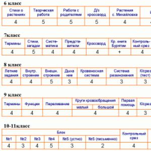
Tests
610-1. Which organisms have a body made up of mycelium?
A) algae
B) bacteria
B) mushrooms
D) protozoa
Answer
610-2. Vegetative propagation in fungi is carried out using
A) dispute
B) gametes
B) mycelium
D) fruiting bodies
Answer
610-3. The fruiting body is characteristic of
A) Bacteria
B) Mushrooms
B) Protozoa
D) Algae
Answer
610-4. The mold fungus penicillium consists of
A) various tissues and organs
B) anucleate cells on which sporangia are located
B) multicellular mycelium and racemose sporangia
D) multicellular mycelium and fruiting body
Answer
610-5. Which of the following representatives belongs to the kingdom of fungi?
A) sphagnum
B) streptococcus
B) penicillium
D) chlorella
Answer
610-6. What fungi do not form mycorrhizae with woody plants?
A) boletus
B) boletus
B) chanterelles
D) tinder fungi
Answer
610-7. Look at the drawing. What letter on it indicates the mycelium?
Answer
610-8. What function does the cap of the fruiting body perform in boletus?
A) serves to attract animals and humans
B) captures solar energy, enabling photosynthesis
B) is the place where spores are formed
D) provides air supply
Answer
610-9. Which of the following fungi does not form mycorrhizae?
A) tinder fungi
B) boletus
B) boletus
D) white
Answer
610-10. What are hyphae?
A) threads that make up the body of the mushroom
B) fungal sporulation organs
B) organs of attachment of the fungus to the substrate
D) photosynthetic part of the lichen
Answer
610-11. Consider a microphotograph of a mukor mold. What is contained in the black balls of this mushroom?
A) nutrients
B) water with mineral salts
B) microscopic spores
D) microscopic seeds
Answer
610-12. Which mushroom is classified as tubular?
A) russula
B) boletus
B) autumn honey fungus
D) champignon
Answer
610-13. What function does the fruiting body of the boletus mushroom perform?
A) structural
B) trophic
B) excretory
D) generative
Answer
610-14. When picking mushrooms, it is important not to damage the mycelium, as it
A) serves as a place for spore formation
B) serves as food for animals living in the soil
B) absorbs nutrients dissolved in water from the soil
D) holds soil lumps together and protects it from erosion
Answer
610-15. Settling on stumps, honey mushrooms use them for
A) attracting pollinating insects
B) obtaining finished organic substances
B) obtaining energy from inorganic substances
D) protection against pathogenic bacteria
Answer
610-16. Why is it often found on a rotten stump? a large number of again?
A) a rotting stump releases heat, which activates the growth of honey mushrooms
B) a rotting stump emits heat, which activates the reproduction of mushrooms
C) honey mushrooms feed on organic matter from dead plants
D) the mycelium of honey mushrooms forms mycorrhiza with the roots of the stump
Answer
610-17. Why are porcini mushrooms often found in oak forests?
A) There is a lot of light in the oak forest.
B) Porcini mushrooms form mycorrhiza with oak roots.
C) Porcini mushrooms have no competitors in the oak forest.
D) In the oak forest there are no animals that feed on porcini mushrooms.
The body of the mushroom is mycelium, consisting of thin threads - gif. The mycelium has a close connection with the substrate, which is due to the osmotic absorption of nutrients. U higher The mycelium of mushrooms is divided into individual cells by partitions - septa, i.e. they have a septate (cellular) mycelium. Inferior mushrooms have noncellular structure mycelium, since its hyphae are not divided into partitions, but are like one branched cell with many nuclei.
Fungi are isolated in their morphophysiological organization from the rest of the world of living beings. They cannot be classified as either plants or animals. There are two theories of the origin of fungi: animal and plant, since fungal cells have characteristics of both animal and plant cells (Table 5.2).
Plant theory fungi suggests their origin from green algae, from which it follows that fungi are primarily a clearly regressive group of plants that have lost chloroplasts.
Animal theory is based on the fact that fungi are initially chlorophyll-free organisms, i.e. come from simple heterotrophic organisms, and not from algae. This theory is preferable, since achlorophyll-free algae, classified as green, accumulate starch as a reserve product, while fungi do not have starch.
Table 5.2. Features of the structure of a mushroom cell
Fungi are heterotrophs. Like bacteria, they are characterized by extracellular digestion, carried out by secreting external environment enzymes. Absorption of broken down nutrients occurs osmotically across the entire surface of the body. Mycelium cells deposit carbohydrates in the form of glycogen, fats in the form of lipid droplets, and proteins in vacuoles as reserve nutrients.
Mushrooms are capable enter into symbiosis with higher plants, forming mycorrhiza(fungus root). Fungi use carbohydrates synthesized by the plant and obtain for it (due to the mineralization of organic compounds) various compounds with nitrogen, phosphorus, and produce growth activators and vitamin-like substances.
Multiply Fungi can be vegetative, asexual and sexual.
Vegetative reproduction can occur by parts of the mycelium (in almost all mushrooms), by budding (yeast). Asexual reproduction occurs due to the formation of zoospores, sporangiospores and conidia.
Zoospores are formed in fungi leading an aquatic lifestyle (chytridiomycetes, oomycetes). Their mobility is ensured by flagella (1 or 2 of them). They are formed inside single-celled zoosporangia and, when ripe, enter the water. They become covered with a shell and grow into a new individual.
Sporangiespores are formed endogenously - inside unicellular sporangia that arise on sporangiospheric hyphae. One sporangium can contain up to 10 thousand spores, which, when ripe, emerge from the sporangium and are distributed by the wind over considerable distances. Once in favorable conditions, the spore grows into a new mycelium (for example, in mucor).
Conidia are formed exogenously on special hyphae - conidiophores. Conidia form chains, detach and, in a favorable environment, germinate into new mycelium (for example, in penicillium).
Sexual reproduction in lower fungi happens:
During the fusion of gametes - gametogamy(isogamy, heterogamy and oogamy);
With the fusion of two multinucleated specialized reproductive organs (gametangia) - zygogamy.
Sexual reproduction in higher fungi:
gametangiogamy; archicarp - female gametangium, antheridium - male (in marsupial fungi);
somatogamy- fusion of haploid somatic cells of heterothallic hyphae (+ and - physiologically different hyphae), for example in higher basidiomycetes.
The sexual process always ends with the formation of a diploid zygote, its meiotic division and sporulation.
The lower fungi include the zygomycota division, the higher ones include the divisions: marsupials, basidiomycota, imperfect.
DEPARTMENT OF ZYGOMYCOTA(ZYGOMYCOTA)
Mucor is widely distributed in nature as white mold (Fig. 5.15). Saprophyte by way of nutrition; develops on soil and food products. Mycelium hyphae are an elongated, overgrown giant cell with many nuclei (non-cellular structure). Nuclei - with a haploid set of chromosomes (n). Numerous vertical sporangiophores with brown-black sporangia develop on the mycelium. As a result of mitosis, the contents of the sporangium break up into many spores (up to 10 thousand). After maturation, the sporangium shell bursts and the spores disperse, germinating into new individuals. Reproduction can be asexual (spores), vegetative (parts of mycelium), and rarely sexual (zygogamy).
With zygogamy (Fig. 5.16), physiologically different hyphae - heterothallic, conventionally designated as + and -, begin to grow towards each other. At the ends of the hyphae, gametangia are formed, separated by septa from the rest of the hyphae. Next, gametangiogamy occurs, consisting in the fusion of 2 specialized reproductive structures (gametangia), not differentiated into gametes, and a zygote with many diploid nuclei is formed. The zygote is covered with a thick brown membrane. After a period of rest, the nuclei undergo meiosis, and the zygote grows into an embryonic sporangium. The haploid nuclei + and - formed after meiosis pass into it. Spores are formed in the sporangium; after they mature, the sporangium is opened, the spores disperse and grow into new mycelia (+ and -).

Rice. 5.15. The structure of mucor (Mucor mucedo): 1 - hypha; 2 - mycelium; 3 - sporangiophore; 4 - sporangium with spores
Some mucor fungi cause mycosis (mucoromycosis) of the lungs (false tuberculosis), brain and other human organs, as well as agricultural plants. Many species of the genus have high enzymatic activity, which is used in the production of “soy cheese” from soybean seeds, alcohol from potato tubers, etc.

Rice. 5.16. Life cycle of mucor (Mucor): A - haploid phase; B - diploid phase: 1 - two heterothallic (opposite in physiological sign) mycelium; 2 - sporangiophore; 3 - sporangium; 4 - disputes; 5 - spore germination; 6 - gametangium; 7 - pendants; 8 - zygospore; 9 - germinating zygospore; 10 - germinating mycelium
DEPARTMENT MARSPIAL FUNGI, OR ASCOMYCOTS(ASCOMYCOTA)
This is one of the most extensive classes of fungi, including more than 30 thousand species. This class includes yeasts, represented by single budding cells, and mushrooms with large fruiting bodies, such as morels and strings. Ascomycotes are widespread in nature in all natural areas. According to their feeding method, they are saprophytes. The mycelium of marsupial fungi is septate, i.e. divided into cells (with a haploid set of chromosomes). A characteristic feature of ascomycots is the presence of bags (asks) formed as a result of the sexual process. Bags are closed structures containing a certain number of ascospores (spores of sexual reproduction) and formed as a result of meiosis.
In many ascomycotas, pouches are formed in the fruiting bodies (subclass Fruit marsupials). There are 3 types of fruiting bodies: cleistothecia, perithecia And apothecium. In other representatives, the bags lie open on the mycelium (subclass Holosumchatae).
Asexual reproduction also plays a large role in the development cycle. Asexual reproduction spores - conidia- are formed as a result of mitosis on mycelium with haploid nuclei (n) or conidiophores of various structures.
The most common and most practical is genus Yeast (Saccharomyces). Yeast is represented by single, oval cells (Fig. 5.17). Yeasts are characterized by vegetative reproduction, carried out by budding; To do this, they need a nutrient medium, the presence of sugar in it and a certain temperature. Under unfavorable conditions it occurs sexual process; When 2 haploid daughter cells fuse (chologamy), a zygote is formed, which turns into a bursa. As a result of meiosis, four spores (ascospores) are formed in the bag, which germinate into new yeast cells.
Baker's yeast (Saccharomyces cerevisiae) combines many cultivated yeasts: alcohol, beer, wine, bakery. All these yeasts decompose sugar into ethyl alcohol and CO 2. So, when yeast is added to the dough, it begins to decompose the glucose present there, formed from starch. In this case, CO 2 is released, which provides the dough with porosity and an increase in volume. When baking, ethanol and CO 2 evaporate.

Rice. 5.17. Brewer's yeast (Saccharomyces cerevisiae): A - unicellular thallus; B - bag with ascospores; B – budding
Yeast is a valuable food and feed product. Contain up to 50% protein, as well as fats and carbohydrates. Synthesized into large quantities vitamins, especially B2. They are used in the treatment of anemia, and also as a source of protein when added to feed products in livestock and poultry farming.
Subclass Fruit marsupials(Carpoascomycetidae)
Representatives of this subclass are characterized by the presence of fruiting bodies containing bags. Fruiting bodies are formed due to a dense plexus of haploid and dikaryon (binucleate) hyphae, also called ascogenous. Fruiting bodies (ascocarps) are of 3 types: closed (closed) - cleistothecia, semi-closed - perithecia, unclosed (open) - apothecia.
The development cycle of ergot proceeds with a change of nuclear phases (Fig. 5.18). Thus, in the fall, cereal plants form sclerotia- dark purple outside and white inside horns, representing the mycelium of the fungus (dehydrated hyphae) in the dormant stage. In winter, sclerotia fall from the cereals onto the soil and overwinter in it. In spring, sclerotia germinate on the soil, forming thread-like outgrowths crowned with heads - stroma. In these stroma, as a result of the sexual process, they are formed fruiting bodies - perithecia, filled with long cylindrical bags (asci) containing filamentous ascospores - spores of sexual reproduction (Fig. 5.19). Spores mature as a result of meiosis during flowering of the cereal. Spores are actively released by the wind, land on the stigma of a flowering cereal and germinate. The resulting mycelium penetrates the ovary of the pistil and destroys it. At the ends of the mycelial hyphae, as a result of mitosis, conidia are released - spores of asexual reproduction, i.e. conidial sporulation occurs. At the same time, the hyphae of the fungus secrete droplets of sweet liquid - “honeydew”. Insects transfer conidia to the flowers of neighboring plants and infect them.

Rice. 5.18. Purple ergot (Claviceps purpurea): A - rye ear with sclerotia (1); B - stroma (2), grown on overwintered sclerotia; B - longitudinal section through the stroma with perithecia; G - longitudinal section through perithecia (3) with bags; D - bag with filamentous ascospores (4); E - conidial sporulation

Rice. 5.19. Development of a bag with ascospores: A, B - formation of a zygote at the apex of the ascogenous hypha; B-E - meiosis and development of the bag with ascospores
Unclosed fruiting bodies - apothecia- found in such representatives as morels (Morchella), stitches (Gyromitra). This open fruiting body is usually saucer-shaped, goblet-shaped, measuring from 0.1 to 10 cm, of various colors - from bright orange or red to brown and black. Upper layer (hymenium) contains many bags. The fruiting bodies of fungi from this group consist of a sterile stalk and a folded or lobed cap (Fig. 5.20).
Morels and lines - edible mushrooms, but when eating the lines, you must first boil them and drain the water.

Rice. 5.20. Ascomycota - appearance and fruiting bodies of morels and strings:
A - conical morel (Morchella coinca); B - common stitch (Gyromitra exculenta); 1 - sections of fruiting bodies
DEPARTMENT OF BASIDIOMYCOTA(BASIDIMYCOTA)
This class combines almost all groups of cap mushrooms, numbering about 30 thousand species. The vegetative body is represented by segmented mycelium, consisting of segmented hyphae.
Reproduction:vegetative(carried out by parts of the mycelium), asexual(conidia) and sexual.
During the sexual process, no special organs of sexual reproduction are formed. The sexual process takes place in the form somatogamy(Fig. 5.21). From the germinating haploid basidiospore, the primary mycelium develops, which then turns into segmented. Each segment is uninucleate. Soon it happens hologamy- fusion of terminal hyphal cells. However, the fusion of the contents of the segments is not accompanied by the fusion of the nuclei. Dikaryons are formed, which then divide synchronously. This is how it is formed secondary dikaryonic mycelium.

Rice. 5.21. Development of basidiomycete fungus. Diagram of the development cycle: A - diagram of the development cycle: 1 - basidium; 2 - basidiospore; 3 - primary mycelium; 4 - dikaryonic mycelium; 5 - fruiting body from dikaryon mycelium; B - development of basidium with basidial spores
A fruiting body is formed on the dikaryon mycelium, which consists of a stump and a cap. Hymenial layer caps can be lamellar or tubular. In the hymenial layer, at the ends of dikaryonic hyphae from 2 nuclear cells, basidia. In their development, basidia are homologous to bursae. The sexual process is completed in the basidium, i.e. The dikaryon nuclei fuse and a diploid nucleus is formed. This single-celled basidia is called Holobasidia. The resulting diploid nucleus is divided by meiosis to form 4 haploid nuclei (see Fig. 5.19, A). By this time, four tubular outgrowths are formed in the upper part of the basidium - sterigmas. The resulting nuclei flow into the sterigmata and 4 basidiospores are formed: 2 conventionally with the sign - and 2 with the sign +. Therefore, the primary mycelia growing from them will be heterothallic. Basidia are formed directly on hyphae or in fruiting bodies various shapes, but more often consisting of a cap and a stalk. The development cycle alternates between 3 phases: haploid(short) are basidiospores, dikaryonny(lasts the main part of life) - dikaryonic mycelium and diploid(short-term) - young basidium before the formation of basidiospores.
DEPARTMENT OF DEUTEROMYCOTA(DEUTEROMYCOTA),OR IMPERFECT MUSHROOMS(FUNGI IMPERFECT!)
Deuteromycots, along with bisidiomycots and ascomycots, are largest group mushrooms, uniting 25-30 thousand species. These fungi are asexual forms (anamorphs) that reproduce asexually - by conidia. Their life cycle takes place in the haploid stage without the sexual process. It is quite possible that deuteromycotes are the most specialized lineages of fungal evolution.

Has great medical importance genus Penicillium. Penicillium has a segmented greenish mycelium consisting of mononuclear segments. The hyphaconidiophores extending upward branch at the upper end onto sterigmas. Latest by appearance resemble a brush or hand and end in a chain of external spores - conidia (Fig. 5.22). Conidia- These are spores of asexual reproduction, formed through mitosis.
A sexual process is also observed, as a result of which closed spherical fruiting bodies of bright yellow color are formed directly on the mycelium - Cleistothecia. Bags with 8 ascospores are formed inside the cleistothecia. Mature ascospores emerge from the bags after rupture of the cleistothecium.
Penicillium (Penicillium), A saprophyte, based on its feeding method, settles on food products and products (fabrics, leather), causing them to spoil. Penicill is used not only for medical practice, but also in the food industry for the preparation of special types of cheese (“Roquefort”).

Rice. 5.22. Deuteromycota penicillium: 1 - mycelium; 2 - conidiophore; 3 - conidia; 4 – sterigmata
The importance of mushrooms in human activity is great. They participate in the cycle of substances in nature. Fungi, like bacteria, mineralize organic matter and take part in the formation of humus. They are used in the food industry for the production of alcohol, wine, beer, kvass, in baking, for the production of proteins and vitamins. Mushrooms form organically active substances- antibiotics, enzymes, organic acids, etc.
Fungi can cause corrosion of metals and destroy leather, paper, and fabrics. Many fungi cause significant harm to humans, animals and plants, causing a number of diseases (mycoses, ringworm, scab), and also lead to food spoilage and thereby cause various poisonings.
LICHEN DEPARTMENT(LICHENES)
This is a group of symbiotropic plants consisting of 2 components - autotrophic algae And heterotrophic fungi. The fungal base of lichens is formed mainly marsupial mushrooms. The algal component consists of species, classified in most cases as representatives of the departments green And blue-green algae. Algae isolated from lichen do not differ from free-living forms. Physiologically, this type of symbiosis is based on intercellular exchange between algae and fungi. The fungus feeds on the algae's carbohydrates, and the algae receive minerals from the fungi. However, symbiosis with fungi leads to the emergence of a new biological quality, which is expressed in the lichen in its ability to reproduce as a single organism.
The vegetative body of lichens is represented by a thallus that has different colors (gray, greenish, brown-brown, yellow or almost black). Morphologically, there are 3 main types of lichen thallus: scale (crust), leafy And bushy(Fig. 5.23), however, there are also transitional forms. The most poorly organized are scale, or cortical, thalli; they have the appearance of powdery, granular, lumpy deposits that grow tightly together with the substrate and do not separate from it without significant damage.

Rice. 5.23. Different types of lichen thalli: A - cortical (graphis - Graphis scripta); B - leafy (xanthoria - Xanthoria); B - bushy (cladonia - Cladonia)
More highly organized lichens have a leafy thallus in the form of plates, scales or rosettes, adhered to the soil or trees with the help of rhizines - analogues of rhizoids, consisting of bundles of fungal hyphae.
The highest organization in their structure is achieved by lichens with a bushy type of thallus, having the appearance of a branched bush (12-15 cm in height) and merging with the substrate only at the base.
According to the anatomical structure, lichens are homeomeric and heteromeric (Fig. 5.24). In the more primitive ones - homeomeric- fungal hyphae and algae are evenly distributed throughout the thickness of the thallus. At heteromeric structure on the cross section of the lichen from above you can see the so-called upper bark. It is formed by intertwining and closely interlocking fungal hyphae. Under the bark, the fungal hyphae lie more loosely, and between them there are algae cells (gonidial layer). Inside the thallus, a core can be distinguished, consisting of loose fungal hyphae and large voids filled with air. Below it is the lower crust, which is similar in structure to the upper crust. Individual hyphae (rhizins) pass through it from the core, securing the lichen in the substrate.
Most lichens tolerate drying out easily. Photosynthesis and nutrition stop at this time, which explains their insignificant annual growth.
Reproduction lichens mainly vegetative, based on the ability of lichens to regenerate from individual areas. It is carried out by fragmentation (separation of sections of the thallus) or with the help of separate groups of algal cells surrounded by fungal hyphae and different in shape - soredia, isidia and lobula (Fig. 5.25). Soredia- the smallest formations of a round shape, including one or several algae cells and surrounded by fungal hyphae. Isidia- tuberculate rod-shaped outgrowths on the upper surface of the thallus.

Rice. 5.24. Anatomical structure of lichen thallus: A - section of the homeomeric lichen thallus: 1 - fungal hyphae; 2 - algal component;
B - section of heteromeric lichen: 1 - upper cortical layer; 2 - gonidial layer; 3 - middle layer with fungal hyphae; 4 - lower cortical layer; 5 - rubbers

Rice. 5.25. Reproduction of lichens: A - soredia; B - isidium
Lobula They look like small scales located vertically on the surface of the thallus or along its edges. In addition, asexual reproduction is observed with the help of spores that are independently formed in both algae and fungi.
Sexual reproduction has not been sufficiently studied, but in general terms it proceeds in the same way as in free-living fungi.
Meaning there are a lot of lichens. They decompose and mineralize soil organic matter. They are pioneers - one of the first to populate rocks, they destroy their surface layer and, dying, form humus on which other plants settle. Lichens are indicators of air purity, as they cannot tolerate even the slightest impurities of sulfur dioxide gases. From some of their types, paint and a special substance - litmus (for the chemical industry) are obtained. In the tundra and forest-tundra, lichens (moss moss) are the main food for deer. Edible lichens are also found in semi-desert and desert regions of Kyrgyzstan and Turkmenistan.
Typical mycelium has the form of thin threads of more or less constant diameter (ranging from 1 to 10 microns, less often 20 microns). In some fungi, such as yeast, the vegetative body is represented by single budding or dividing cells. If such budding cells do not disperse, a pseudomycelium. In the process of evolution, fungi have formed various structures that perform one or another adaptive function. Thus, in mucor fungi, aerial arcuate hyphae are formed - stolons. With their help, the fungus quickly spreads across the substrate. In places of contact with it, they form rhizoids- bundles of short, branched, root-like hyphae that perform the function of attachment.
Intensively branching hyphae during growth often form among themselves anastomoses– short cross bridges. With abundant development of anastomoses, the mycelium takes the form of a mesh and becomes more durable. Often anastomoses develop due to lack of nutrition. Through them, metabolism occurs and, most importantly, migration of nuclei from one hypha to another occurs.
Basidial fungi are characterized by the formation buckles. These are small cells that are located on the side of the hyphae opposite the transverse partitions. They act as a duct for moving one of the dikaryon nuclei from the upper cell to the lower one.
In some predatory fungi, in the presence of small invertebrates, such as nematodes, sticky loops, passive and compression rings and other types of fishing devices.
Often, fungal mycelium breaks up into individual thin-walled cells of various shapes - oidia. Often their formation is associated with the onset of unfavorable conditions. Under suitable conditions, the oidia germinate into new mycelium. In approximately the same way, thick-walled mycelium with a large supply of nutrients is formed on vegetative mycelium. chlamydospores, however, unlike oidia, during the process of formation they form their own dark-colored shell under the old hyphal shell. Chlamydospores can withstand drying and other adverse environmental factors and can remain viable for up to 10 years. They are found in many mushrooms. In some (for example, smut) chlamydospores are an obligatory stage in the life cycle, in others they are formed when the nutrient substrate is depleted and other unfavorable conditions (fungi from the genera Fusarium, Alternaria, Helminthosporium, Phytophthora and etc.).
Many fungi are characterized by various formations consisting of interlacing hyphae - rhizomorphs, strands, mycelial films, stroma, sclerotia. Rhizomorphs They are powerful dark-colored branched cords up to several meters long, consisting of parallel and anastomosing hyphae. They serve for the spread of fungi, conduction of nutrients, and reproduction. Well known, for example, are the rhizomorphs of honey mushroom ( Armillaria mellea), thanks to which the fungus quickly spreads along the tree trunk and can move to another tree.
Mycelial strands formed by a relatively small number of parallel hyphae, which are glued together by mucous walls or connected by short anastomoses. In some species of fungi, the outer elements form a cortex of thin, durable, dark-colored hyphae, and the inner elements form a core of wider, colorless hyphae. This type of strands is characteristic of the dangerous wood-destroying house fungus ( Serpula lacrymans).
Mycelial films They are a layer of tightly intertwined hyphae located in different directions. They can be from a few millimeters to half a centimeter thick; form on the surface of the substrate or in cracks in the bark of trees. Often found in tinder fungi.
Many mushrooms form mycelial stroma- fleshy or woody plexuses of hyphae penetrating the substrate. Fruiting bodies or other sporulation organs are formed on the surface or inside such plexuses. Stromas can be of various shapes and colors. Mycelial stroma is characteristic of many ascomycetes.
A very common formation consisting of tightly woven dehydrated hyphae are sclerotia. Their sizes range from microscopically small to 20–30 cm in diameter. They are varied in shape and rich in reserve nutrients. The main function of sclerotia is to endure unfavorable conditions for a long time and preserve the individual (species). The inner part of the sclerotium usually consists of colorless hyphae, the outer part of dark-colored thick-walled hyphae. There are three types of sclerotia: the first includes sclerotia, which consist exclusively of a plexus of hyphae (for example, in fungi from the genera Claviceps, Botrytis, Sclerotinia, Typhula and etc.); The second type includes sclerotia, which are often called mummies - not only fungal hyphae take part in their formation, but also host tissues (for example, mummified apples, infected Monilinia fructigena; insect larvae affected by species of the genus Cordiceps). The third type is called pseudosclerotia or microsclerotia. They consist of thick-walled, colored hyphae, usually formed inside the tissues of affected plants or on the hyphae of the mycelium during the cultivation of the fungus.
Sclerotia can persist for a very long time and then, under favorable conditions, germinate, usually forming sporulation organs.
In many higher fungi, at certain stages of the life cycle, a kind of tissue is formed from intertwined hyphae - plectenchyma. This is a plexus and fusion of hyphae, each of which grows independently, independently of the others. Plectenchyma is called false tissue, since the cells of the mycelium threads that make it up divide only in one direction, and not in different directions, as in plants. Based on their structure, there are two types of plectenchyma: paraplectenchyma And prosoplectenchyma. Paraplectenchyma is represented by isodiametric cells and externally resembles plant parenchyma. Prosoplectenchyma is formed by elongated cells arranged more loosely than paraplectenchyma. The fruiting bodies of mushrooms and other structures are formed from false tissues.